Growth hormone after traumatic brain injury: what a functional MRI endpoint can show
Functional MRI can record a signal related to brain activity under defined conditions.
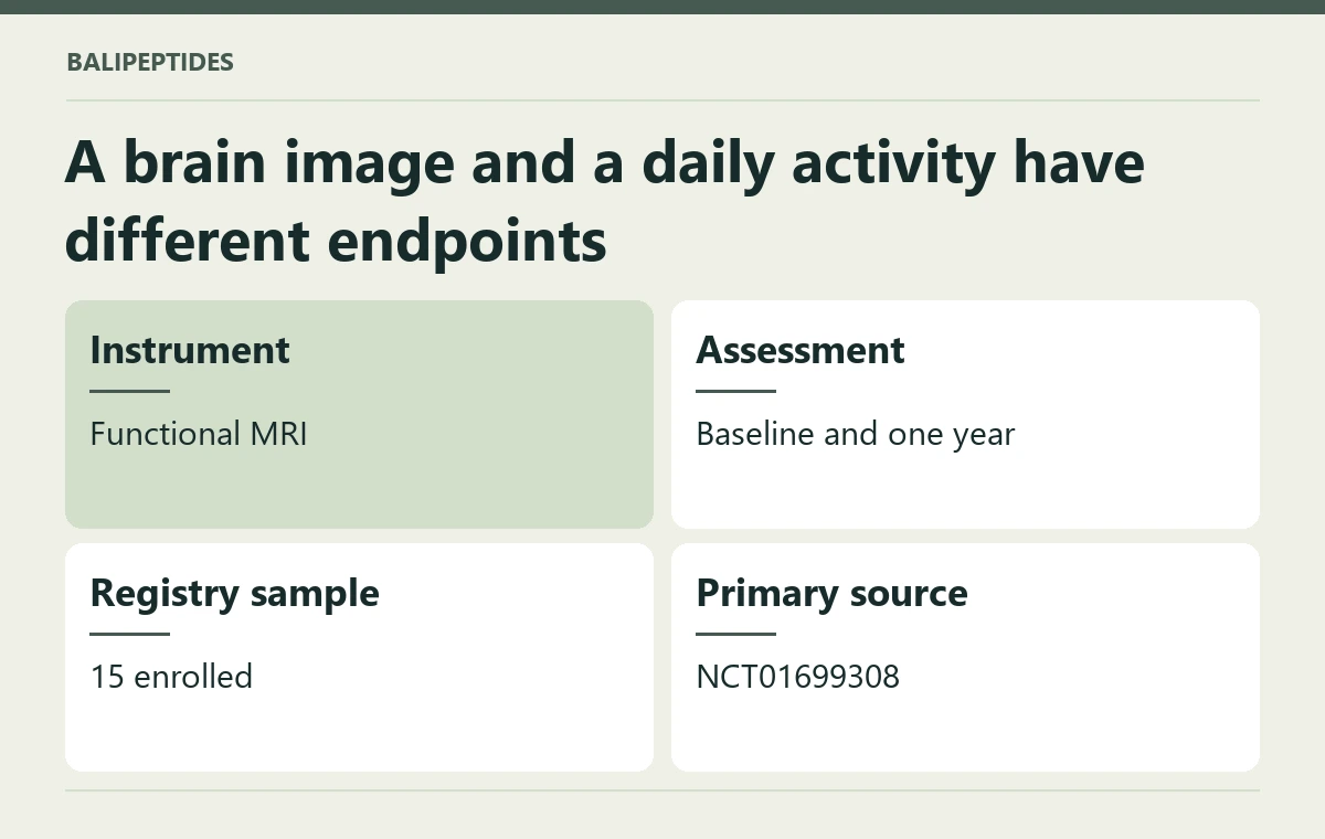
Functional MRI can record a signal related to brain activity under defined conditions. A growth hormone study after traumatic brain injury registered a change in brain function assessed by functional MRI. A scan outcome is valuable, but it does not by itself describe a person's return to daily activities.
A brain image and a daily activity have different endpoints
The protocol compares measurements at baseline and one year. A follow-up scan can ask about brain function in the studied group; it does not substitute for an assessment of symptoms, daily tasks or the individual's hormonal diagnosis. An imaging difference can be scientifically interesting even when its practical meaning remains open. Readers should check the task performed during scanning, the brain region analyzed and how any reported outcome was compared before using it to support a broader recovery claim.
The method determines what a tissue number means
A brain region showing a different signal during a task is a finding about the scan and the task performed. Whether someone can follow a conversation or keep up at work calls for another kind of measurement. Identify which cognitive or everyday function, if any, the protocol assessed directly alongside the imaging.
Functional imaging and everyday experience can diverge. A participant could show a changed imaging signal but still require help planning daily tasks. Conversely, a person could develop useful coping strategies without a dramatic change in a particular image. The way to connect these questions is to measure the functional outcome independently and analyze its relationship with the imaging result.
Participant and measurement details
- People in the record: 15 (actual enrollment). Eligible ages: 18 years to 55 years.
- Clinical setting: Traumatic Brain Injury.
- Named study interventions: Genotropin.
- Study design: phase 1, interventional, non randomized, parallel.
- Measured here: Change in brain function assessed by functional MRI.
- Research locations listed in the record include: Lexington, United States.
- Study calendar in the registration: start 2009-12-01; primary completion 2014-04-01.
- Primary record: Joe Springer. ClinicalTrials.gov, NCT01699308.
Someone in Bali consulting a specialist after brain injury can bring the original scan report and any standardized functional assessments. The Somatropin product listing serves a different purpose. It cannot establish a hormone diagnosis or identify what a change in one scan means for recovery.
